Exploring the Intriguing World of Sheep Eyeball: A Comprehensive Guide
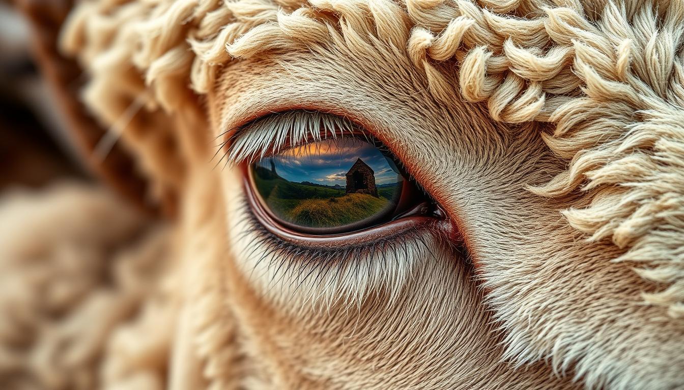
When you explore sheep eyeball anatomy, you find that sheep have been key in biomedical research. This has been especially true in the 1970s and continues today1. This study offers a chance to dive into the details of sheep eye anatomy. You can learn more about animal anatomy at petpawza.
Sheep eyeballs are great for studying eye anatomy because they are similar to human eyes. This makes them very useful for teaching1. The sclera, a key part of the human eyeball, is also found in sheep eyeballs. Its thickness changes depending on where it is on the eyeball2. Knowing about sheep eyeball anatomy helps us understand the eye better, which is important for biology and anatomy students.
Key Takeaways
- You will learn about the external features of the sheep eyeball, including the sclera and episclera.
- The internal structure of the sheep eyeball, including the choroid and retina, will be explored in detail.
- Sheep eyeballs are used in biomedical research due to their similarities to human eyes1.
- The scleral stroma contains collagen-rich extracellular matrix (ECM) and bundles of parallel-aligned collagen fibrils2.
- Understanding sheep eyeball anatomy can provide valuable insights into the structure and function of the eye.
- Sheep eyeballs are an excellent model for studying eye anatomy and are used in educational settings1.
Understanding the Importance of Sheep Eyeball Study
Studying sheep eyeballs is key to understanding the eye’s details. The importance of continuing professional development in vet science shows the need for hands-on learning. Sheep eye dissection is a great way to improve animal care skills. This is especially true for sheep eye structure, which is very similar to the human eye, making it perfect for study3.
There are many benefits to studying sheep eyeballs. It’s a cost-effective choice for educational purposes3. The similarities between sheep and human eyes make it a great model for studying eye functions. By looking at the sheep eye structure, we can learn a lot about eye anatomy and physiology. This knowledge can help improve human eye care and treatment4.
Studying sheep eyeballs also gives us insights into sheep’s visual perception and behavior. This is useful in agriculture and vet care5. By understanding how sheep see and react, farmers and vets can better care for them. Overall, studying sheep eyeballs is both fascinating and rewarding. It offers valuable insights into sheep biology and eye anatomy4.
External Features of the Sheep Eyeball
The sheep eyeball has three main parts: the cornea, sclera, and conjunctiva6. These parts are key to the eye’s function. The cornea is clear and at the front. The sclera is white and tough, protecting the eye6. The conjunctiva is thin and keeps the eye moist.
Some cool sheep eye facts are that it has a tapetum lucidum and four eye-moving muscles6. Humans have six. The sheep eye function is special because its sclera is tough, making it easy to dissect6.
Looking at the sheep eyeball’s parts, we see how they help it see7. Its features are similar to ours, making it great for learning8.
| External Feature | Description |
|---|---|
| Cornea | Transparent layer on the front of the eye |
| Sclera | White, tough layer that provides protection |
| Conjunctiva | Thin membrane that covers the eye and helps to keep it moist |
Complete Sheep Eyeball Anatomy: Layer by Layer
Understanding the sheep eyeball’s anatomy is key in fields like veterinary science and biology. It has several layers, each with its own role. You can learn more about the sheep eye anatomy to understand its structure better.
The outermost layer is the sclera, a tough, white, and fibrous layer that protects the eye9. The middle layer is the choroid, a vascular layer that supplies blood to the eye10. The innermost layer is the retina, a complex layer that detects light and sends visual signals to the brain11.
It’s important to compare sheep eyes with human eyes when studying the sheep eyeball. For example, sheep have four extrinsic eye muscles, while humans have six10. The vitreous humor, a semi-fluid substance, fills the central cavity of the sheep eye, helping to maintain its shape10.
To understand the sheep eyeball anatomy better, look at the following key components:
- The retina, responsible for light detection and visual signal transmission
- The choroid, supplying blood to the eye
- The sclera, providing protection to the eye
Studying the sheep eyeball’s anatomy helps us understand the eye’s complex structures and functions. This is crucial for fields like veterinary science and biology9. The study of the sheep eyeball is fascinating and offers valuable insights into the eye’s anatomy and function11.
Essential Components of Sheep Eye Structure
Exploring the sheep eye structure reveals its complex makeup. The lens, iris, and optic nerve are key parts that help us see. The first web source, sheep eye dissection, is a cost-effective way to study these parts, unlike cow eye dissection12.
The sheep eye structure has layers like the sclera, choroid, and retina. The optic nerve, about 3 mm thick13, is vital for sending visual info to the brain. The vitreous humor, a clear gel, fills the space between the lens and retina, keeping the eye’s shape13.
To really understand the sheep eye structure, let’s look at its parts:
- Lens: focuses light onto the retina
- Iris: controls the amount of light entering the eye
- Optic nerve: transmits visual information to the brain
- Sclera: provides protection and structure to the eye
- Choroid: supplies blood and oxygen to the retina
By dissecting the sheep eye structure, you learn how these parts work together for vision12. This knowledge is useful in biology, medicine, and veterinary science, making it a valuable learning experience14.
The Fascinating Process of Sheep Eye Dissection
Sheep eye dissection is a cool way to learn about the eye’s anatomy and sheep eye function. First, get a sheep eyeball from a local farm or butcher shop, keeping ethics in mind15.
It’s key to know the sheep eyeball’s anatomy before you start. This includes its similarities and differences to human eyes16.
To set up, you’ll need tools like a sharp scalpel, forceps, a cutting board, and gloves15.
Following a step-by-step guide and wearing gloves are crucial for a safe dissection. Make sure a parent is there to help15.
As you delve into the sheep eye facts, you’ll discover how vision works. You’ll see how these mechanisms help the eye function properly.
For more details on sheep eyeball dissection, check out
By following these steps and taking safety seriously, you’ll understand the sheep eye better. You’ll learn about its sheep eye facts and sheep eye function17.
How Sheep Vision Works
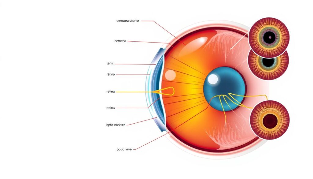
Sheep vision is a complex process that involves the sheep eyeball and its various components. The sheep eye anatomy is designed to provide a wide field of vision. This is essential for detecting predators.
According to18, sheep have a panoramic field of vision ranging from 320-340 degrees. But their binocular field is limited to only 20-60 degrees.
The light processing mechanism in sheep vision involves the absorption of light by the retina. This light is then transmitted to the brain for interpretation. This process is crucial for sheep to navigate their environment and detect potential threats.
As noted in19, the control of retinal illumination in different light environments is essential. This allows sheep to adapt to their surroundings.
Some key features of sheep vision include:
* A wide field of vision
* Limited binocular vision
* Ability to detect colors, except for red due to red-green colorblindness18
* Strong visual memory capabilities, allowing them to recollect up to 50 unique human or sheep faces for up to 2 years18
Understanding how sheep vision works is essential. It helps us appreciate the complexities of sheep eye anatomy and its role in the animal’s overall behavior and survival. By studying the sheep eyeball and its components, we can gain a deeper appreciation for the intricate processes that occur in the natural world20.
| Feature | Description |
|---|---|
| Panoramic field of vision | 320-340 degrees |
| Binocular field | 20-60 degrees |
| Color vision | Ability to detect colors, except for red |
Comparing Sheep Eyes to Other Species
Understanding sheep eye dissection means knowing the similarities and differences with other species’ eyes. The sheep eye structure is similar to human eyes in many ways, but also has its own unique features21. For example, sheep corneas are thicker than those of alpacas, showing a difference in eye anatomy22.
A study on local nondescript sheep showed how their eyes change with age. The study found that some parts of the eye grow, but not as much as others23. This helps us understand how sheep eyes develop and change over time. The sheep eye structure also has special measurements like axial globe length and lens thickness21.
Sheep eyes are different from others, like having a thicker cornea and a deeper anterior chamber22. These differences come from each species’ unique evolutionary history. By studying sheep eye dissection and sheep eye structure, researchers can learn more about eye anatomy and its variations.
Modern Applications in Veterinary Science
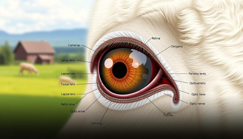
Sheep eye facts and function are key in veterinary science. They help with diagnosis, treatment, and research24. Studying sheep eyes has helped us understand diseases like ovine infectious keratoconjunctivitis (OIKC)25. This has led to better ways to diagnose and treat these conditions, helping sheep stay healthy.
Tools like ocular swabs are used to check for eye diseases in sheep25. The SRUC Veterinary Services sees more of these swabs during certain times25. Tests for M. conjunctivae PCR cost £34.50, and ovine mycoplasma DGGE is £95.7025.
The table below shows some diagnostic tools and treatments used in veterinary science:
| Diagnostic Tool | Price |
|---|---|
| M. conjunctivae PCR | £34.50 |
| Ovine mycoplasma DGGE | £95.70 |
| Antibiotic sensitivity testing | £35.35 |
Knowing about sheep eyes is crucial for veterinary science24. By studying sheep eyes, researchers can find new ways to treat diseases. This helps keep sheep healthy and improves their quality of life.
Conclusion: The Significance of Understanding Sheep Eye Biology
Exploring sheep eyeballs reveals the importance of studying their biology26. The dissection of sheep eyes is a key early science lesson. It teaches students about anatomy and how we see the world26. Although it might smell, it makes learning more engaging and shows the variety of life forms26.
Knowing about sheep eye biology also helps in veterinary science26. Sheep eyes, with their special features like the tapetum lucidum, help us understand how different animals see. This knowledge can lead to better ways to diagnose and treat animals and humans27. Studying sheep eyes alongside human eyes can also help us learn more about our own vision and how to improve it26.
As you move forward in science, remember the value of sharing knowledge and sparking curiosity26. Teachers and family members are crucial in shaping future scientists and doctors26. By exploring sheep eye biology, you can open up new areas of scientific research. This can have a big impact on our world.
FAQ
Why do scientists choose sheep eyes for research?
How are sheep eyes similar to human eyes?
What is the educational value of studying sheep eyeballs?
What are the external features of the sheep eyeball?
Can you describe the internal structure of the sheep eyeball?
What are the essential components of sheep eye structure?
How do I safely dissect a sheep eyeball?
How does sheep vision work?
How do sheep eyes compare to other species?
How are sheep eyeballs used in modern veterinary science?
Source Links
- https://www.mdpi.com/2079-7737/11/9/1251 – The Sheep as a Large Animal Model for the Investigation and Treatment of Human Disorders
- https://pmc.ncbi.nlm.nih.gov/articles/PMC7187923/ – Scleral structure and biomechanics – PMC
- https://www.homesciencetools.com/product/sheep-eye-specimen/?srsltid=AfmBOopHqOKx-elIJxjkddlg3Bfw-Y892Xw9VsmDzC2wZt0LPnVT5Iws – Sheep Eye Specimen
- https://www.cambridge.org/core/journals/animal-science/article/eye-of-the-domesticated-sheep-with-implications-for-vision/9EFB8E9C4B51740ECCFA120400269E02 – The eye of the domesticated sheep with implications for vision | Animal Science | Cambridge Core
- https://www.nfacc.ca/resources/codes-of-practice/sheep/appendix_e.pdf – Code of Practice for the Care and Handling of Sheep
- https://gems.education.purdue.edu/wp-content/uploads/2019/01/Sheep_Eye_Dissection.pdf – SHEEP EYE DISSECTION PROCEDURES
- https://tra.extension.colostate.edu/wp-content/uploads/sites/9/2017/05/17.Sheep_Eye.pdf – PDF
- https://www.homesciencetools.com/product/sheep-eye-specimen/?srsltid=AfmBOoqp48Q_J0Hnmsc8hFc1gntfBiFC7rqtu309AQjb4dDl6Rtc_jIF – Sheep Eye Specimen
- http://www.mrromswinckel.com/uploads/1/1/7/6/1176577/sheep_eye_dissection_lab_handout.pdf – PDF
- https://bettereducate.com/uploads/file/41963_01SampleSheepEyeDissection_01SampleSheepEyeDissection.pdf – Microsoft Word – sheep eye dissection lab-1.doc
- https://www.homesciencetools.com/product/sheep-eye-specimen/?srsltid=AfmBOooojzqOPmtaoKkfBlj38Qq5sS5BeLixkOnU2G1FUgPjKFN-I_Hs – Sheep Eye Specimen
- https://www.homesciencetools.com/product/sheep-eye-specimen/?srsltid=AfmBOopCvJ1k4aFZPmQ9lUAG6WipnMsHo9_B9sK_cSW5_sTHpojUu4Gq – Sheep Eye Specimen
- https://www.wlwv.k12.or.us/cms/lib/OR01001812/Centricity/Domain/1341/LAB – Dissection of the Sheep Eye.pdf – Microsoft Word – LAB – Dissection of the Sheep Eye
- https://www.mrgscience.com/uploads/2/0/7/9/20796234/activity_21__sheep_eye_dissection.pdf – PDF
- https://pineknollsheepandwool.com/sheep-eyeball-for-homeschoolers/ – Sheep Eyeball for Homeschoolers – Pine Knoll Sheep & Wool
- https://curiositykilledthecation.wordpress.com/2018/09/17/dissection-eye/ – Dissection : A Tour of the Eye
- https://middleschoolscience.com/2016/07/13/sheep-head-dissection-brains-tongues-and-eyes/ – Sheep Head Dissection – Brains, Tongues, and Eyes
- https://archives.evergreen.edu/webpages/curricular/2011-2012/m2o1112/web/sheep.html – ARCHIVE – Goats/Sheep – Comparative Physiology of Vision
- https://pmc.ncbi.nlm.nih.gov/articles/PMC4643806/ – Why do animal eyes have pupils of different shapes?
- https://www.homesciencetools.com/product/sheep-eye-specimen/?srsltid=AfmBOopEW77SEOdxFmH6kmZbLMeIkakWlrYI_6ocd_jm2P30lS7r5Zr7 – Sheep Eye Specimen
- https://pmc.ncbi.nlm.nih.gov/articles/PMC4935764/ – The eye of the Barbary sheep or aoudad (Ammotragus lervia): reference values for selected ophthalmic diagnostic tests, morphologic and biometric observations
- https://avmajournals.avma.org/view/journals/ajvr/78/1/ajvr.78.1.80.xml – No title found
- https://journals.tubitak.gov.tr/cgi/viewcontent.cgi?article=1079&context=veterinary – Ocular ultrasonography and echobiometry in sheep
- https://pmc.ncbi.nlm.nih.gov/articles/PMC7151925/ – Immunoassay Applications in Veterinary Diagnostics
- https://www.sruc.ac.uk/veterinary-surveillance-blog/ocular-disease-investigation-in-sheep/ – Ocular disease investigation in sheep
- https://www.cram.com/essay/Science-Experience-Essay/F36VYJLGREE5 – My Reflection On The Dissection Of Sheep Eyeballs
- https://www.ncbi.nlm.nih.gov/pmc/articles/PMC7311881/ – Ophthalmic findings in sheep treated with closantel in Curitiba, Brazil


